Journal of Clinical and Medical Images (ISSN 2640-9615)

Volume 8

Improvement of the Quality of Prostate Cancer Diagnosis Using AI In Digital Pathology Short Heading: Quality Improvement by Using AI In Pathology

1. Abstract 1.1. Aim Automated AI-based tools are becoming increasingly important in modern medicine, including pathology, by significantly supporting pathologists’ diagnoses and reducing human biases. Histopathological evaluation of prostate biopsies plays a crucial role in diagnosing prostate cancer.

Improvement of the Quality of Prostate Cancer Diagnosis Using AI In Digital Pathology Short Heading: Quality Improvement by Using AI In Pathology Read More »

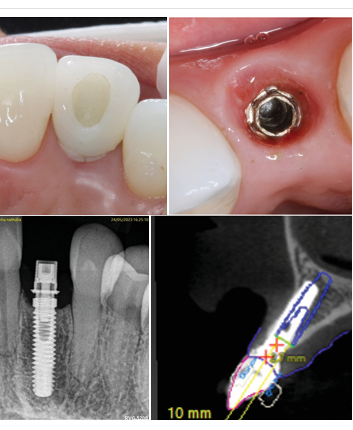
Guided Bone Regeneration of a Seibert Class III Defect with Bioactive Calcium Phosphosilicate Bone Graft (Tent Pole Technique) after Implant Failure: Human Histomorphometry and Clinical Report

1. Abstract This clinical case aimed to achieve two main objectives. The first was to determine if the use of only calcium phosphosilicate bone graft as a regeneration material (with no autologous bone added), on a severe vertical and horizontal mandibular defect (created by an implant failure), would allow enough bone to be obtained to

Guided Bone Regeneration of a Seibert Class III Defect with Bioactive Calcium Phosphosilicate Bone Graft (Tent Pole Technique) after Implant Failure: Human Histomorphometry and Clinical Report Read More »

Use of Optical Coherence Tomography images to Differentiate Between Normal Skin, Skin Lesions, and Melanoma: A Pilot Study

1. Abstract Optical coherence tomography is a technique used extensively in ophthalmology to diagnose several ocular diseases. It is being used in dermatology to diagnose skin conditions.

Use of Optical Coherence Tomography images to Differentiate Between Normal Skin, Skin Lesions, and Melanoma: A Pilot Study Read More »